16 Performing FAST
Lo Zhen Zhen
General Points
- Utility: FAST and eFAST are primarily used in trauma settings, allowing for immediate detection of life-threatening conditions.
- Speed: They can be performed quickly, making them ideal for emergency scenarios.
- Non-Invasive: As ultrasound-based techniques, they are non-invasive and can be repeated as needed.
Indications for a FAST Scan in Trauma Care
The FAST scan is primarily indicated in the evaluation of blunt or penetrating abdominal trauma. It is crucial in the rapid assessment of patients with suspected internal bleeding, assessing for the presence of free fluid in the abdominal, pelvis or pericardial cavities, which may indicate organ injury or hemoperitoneum.
Indications
- Trauma: Any patient with blunt or penetrating trauma.
- Instability: Hemodynamically unstable patients where the source of instability is unclear.
- Monitoring: In certain scenarios, to monitor known fluid collections.
- Other clinical cases: FAST is also useful in non-trauma situations such as in cases of ruptured ectopic, ruptured ovarian cyst and ascites.
Detailed Guide on Probe Selection, Placement, and Movement Techniques
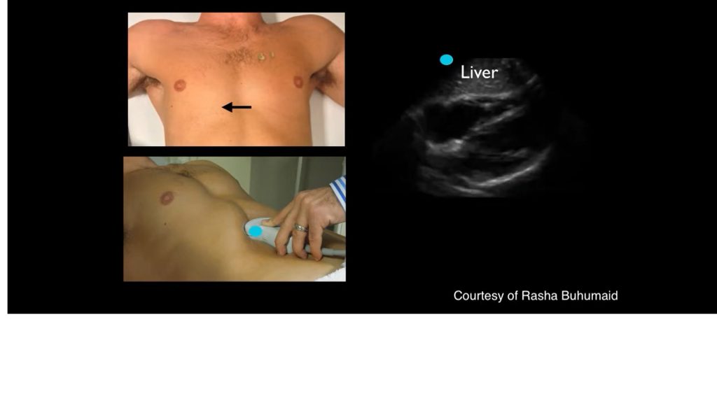
- Probe Selection: The curvilinear (low frequency) probe is typically used due to its deeper penetration, which is ideal for abdominal scanning.

Image showing all 4 quadrants involved in FAST Scan. Adapted from “Fast Protocol in Emergency Medicine – Radiology” by Kashmiri Medicos is licensed under CC BY 4.0
- Probe Placement and Orientation:
- Right Upper Quadrant (RUQ): Place the probe in the mid-axillary line, just below the costal margin. The probe marker should point towards the patient’s head. This view assesses the hepatorenal space (Morison’s pouch) for free fluid.
Normal Right Upper Quadrant study in FAST scan. Adapted from “RUQ efast normal examination” by International Emergency Medicine Education Project is licensed under CC BY 4.0
-
-
- Left Upper Quadrant (LUQ): Position the probe at the posterior axillary line, just below the costal margin, with the probe marker pointing cephalad. This view evaluates the splenorenal space for fluid accumulation.
-
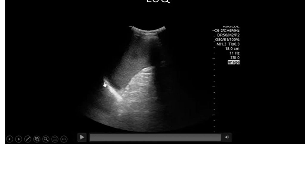
Image showing Left Upper Quadrant in FAST Scan. Adapted from “POCUS Cases 9: Abdominal Free Fluid” by EM Cases is licensed under CC BY 4.0
-
-
- Suprapubic: Position the probe above the pubic symphysis with the marker oriented to the patient’s right side. This view is for assessing the pelvis, particularly the rectovesical pouch in males and the rectouterine pouch (Pouch of Douglas) in females.
-
Normal Scan for Suprapubic window in a Male Patient during FAST Scan. Adapted from “efast normal male pelvic longitudinal view” by International Emergency Medicine Education Project is licensed under CC BY 4.0
Normal Scan for Suprapubic window in a Female Patient during FAST Scan. Adapted from “efast normal female pelvic longitudinal view” by International Emergency Medicine Education Project is licensed under CC BY 4.0
-
-
- Subxiphoid: Place the probe below the xiphoid process with the marker pointing to the patient’s right. This view assesses the pericardial space for fluid.
-
Subxiphoid window view in FAST scan. Adapted from “Video 1 Tutorial on basic echocardiography views” by International Emergency Medicine Education Project is licensed under CC BY 4.0
- Movement Techniques: Gentle, sweeping motions are required to fully visualize each area. Adjust the depth and gain settings on the ultrasound machine to optimize the image.
Anatomy Landmark Summary and Structures Identified
- RUQ: Focus on Morison’s pouch between the liver and right kidney. Look for free fluid.
- LUQ: Examine the splenorenal recess for fluid.
- Subxiphoid: Assess the pericardial space for fluid indicating cardiac tamponade.
- Suprapubic: Evaluate the pelvis, especially the recto-uterine or recto-vesical pouch.
- Thoracic (eFAST): Check for pneumothorax (absence of lung sliding) and hemothorax (fluid in pleural space)
Interpretation of Sonographic Findings
- Normal Findings: The absence of free fluid in the hepatorenal space, splenorenal space, pericardial space, and pelvis.
- Pathological Conditions: The presence of anechoic (black) areas in these spaces indicates free fluid, suggestive of internal bleeding. In the subxiphoid view, fluid around the heart indicates a pericardial effusion, which may be due to a cardiac tamponade in trauma settings.
Free fluid at Right Upper Quadrant View. Adapted from “SS Video 12 Abnormal Right Upper Quadrant” by International Emergency Medicine Education Project is licensed under CC BY 4.0
Free Fluid at Left Upper Quadrant. Adapted from “SS Video 16 Abnormal Left Upper Quadrant” by International Emergency Medicine Education Project is licensed under CC BY 4.0
Large Pericardial Effusion seen at Subxiphoid Window in FAST Scan. Adapted from “SS Video 2 Large Pericardial Effusion” by International Emergency Medicine Education Project is licensed under CC BY 4.0
Free fluids retrovesically in a Transverse Suprapubic View in FAST Scan. Adapted from “SS Video 19 Abnormal Transverse Pelvic View” by International Emergency Medicine Education Project is licensed under CC BY 4.0
Free fluids seen in longitudinal suprapubic window in FAST Scan. Adapted from “SS Video 20 Abnormal Pelvis View” by International Emergency Medicine Education Project is licensed under CC BY 4.0
