24 Identifying and Obtaining Standard Echocardiographic Views
Mexmollen Marcus
In cardiac POCUS, there are several standard views that are essential for a comprehensive evaluation of the heart. Here’s how to obtain each and what they typically show:
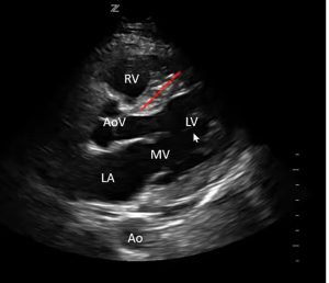
Parasternal Long Axis (PLAX) View:
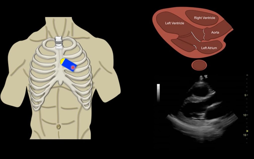
- How to Obtain: Place the transducer just left of the sternum, in the third or fourth intercostal space, with the indicator pointing towards the patient’s right shoulder.
- Visible Structures: Left ventricle, right ventricle, aortic valve, mitral valve, left atrium, and part of the aorta.
- Clinical Relevance: Ideal for assessing left ventricular function, wall motion, and valvular abnormalities, particularly of the mitral and aortic valves.
Parasternal Short Axis (PSAX) View:
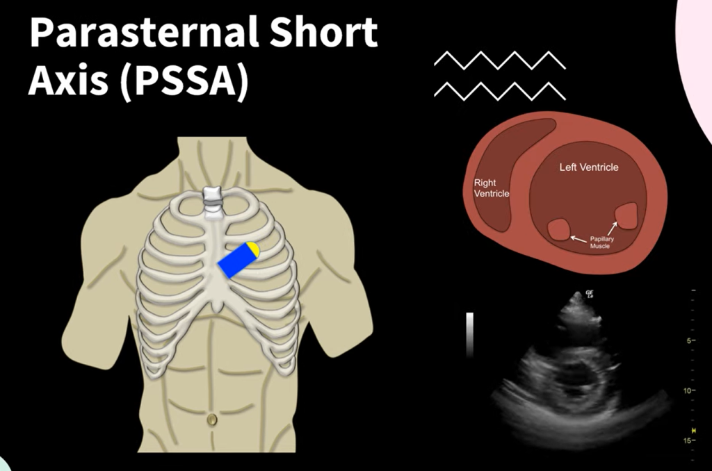
- How to Obtain: Rotate the transducer 90 degrees clockwise from the PLAX position, with the indicator pointing towards the left shoulder.
- Visible Structures: Cross-sectional view of the left ventricle, right ventricle, mitral valve, tricuspid valve, and papillary muscles.
- Clinical Relevance: Useful for evaluating the size and function of the left and right ventricles, and observing the motion of the heart walls.
Apical Four-Chamber View:
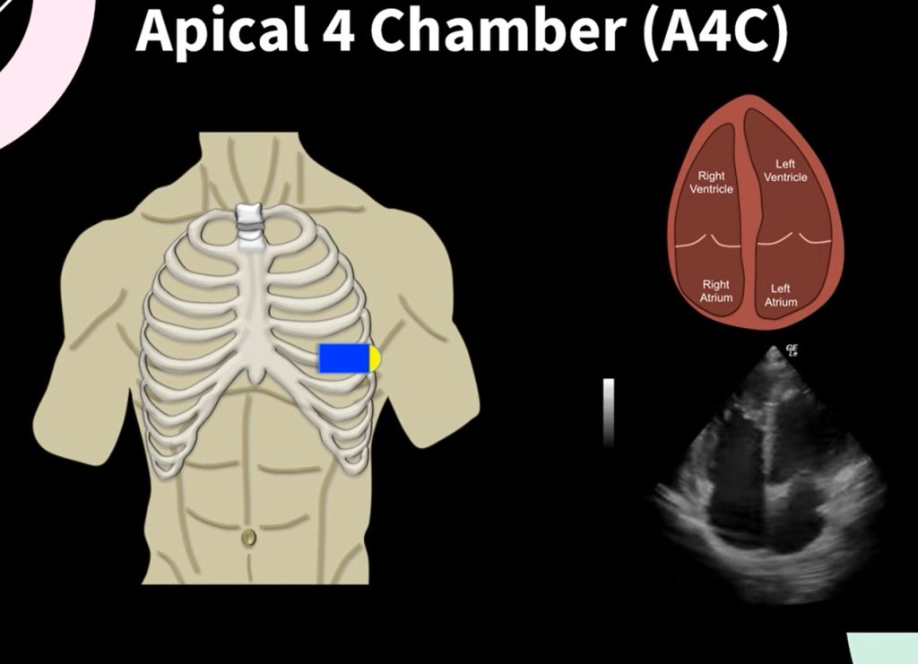
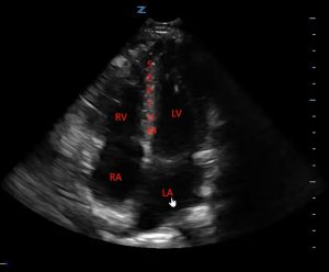
- How to Obtain: Place the transducer at the point of maximal impulse (PMI), typically near the apex of the heart, with the indicator pointing towards the left axilla.
- Visible Structures: Left and right atria, left and right ventricles, tricuspid valve, and mitral valve.
- Clinical Relevance: This view allows for the assessment of chamber sizes, valvular function, and overall cardiac function.
Subcostal View:
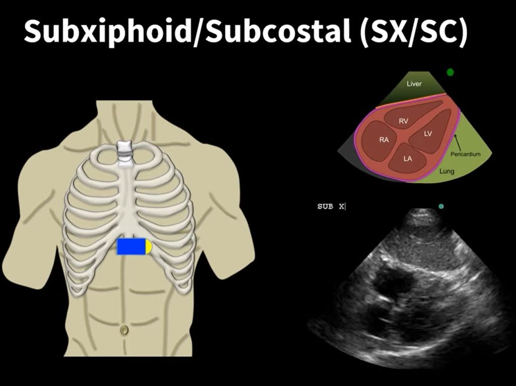
- How to Obtain: Position the transducer just below the xiphoid process, pointing upwards under the rib cage, with the indicator pointing to the patient’s right side.
- Visible Structures: All four heart chambers, inferior vena cava (IVC), and descending aorta.
- Clinical Relevance: Particularly useful in patients with lung disease where air in the lungs may obstruct other views. It’s also ideal for assessing IVC size and collapsibility to estimate right atrial pressures.
IVC View:
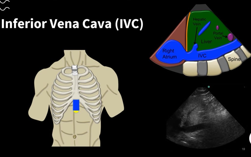
- How to Obtain: From the subcostal position, angle the transducer to focus on the IVC entering the right atrium.
- Visible Structures: Inferior vena cava and its entry into the right atrium.
- Clinical Relevance: Useful for assessing fluid status and right atrial pressure based on the diameter and respiratory variation of the IVC.
Key Structures and Their Clinical Relevance
Each of these views provides crucial information:
- Heart Chambers: Evaluate the size and function.
- Heart Valves: Look for structural abnormalities or malfunctions.
- IVC and Aorta: Assess for signs of volume status and systemic circulation issues.
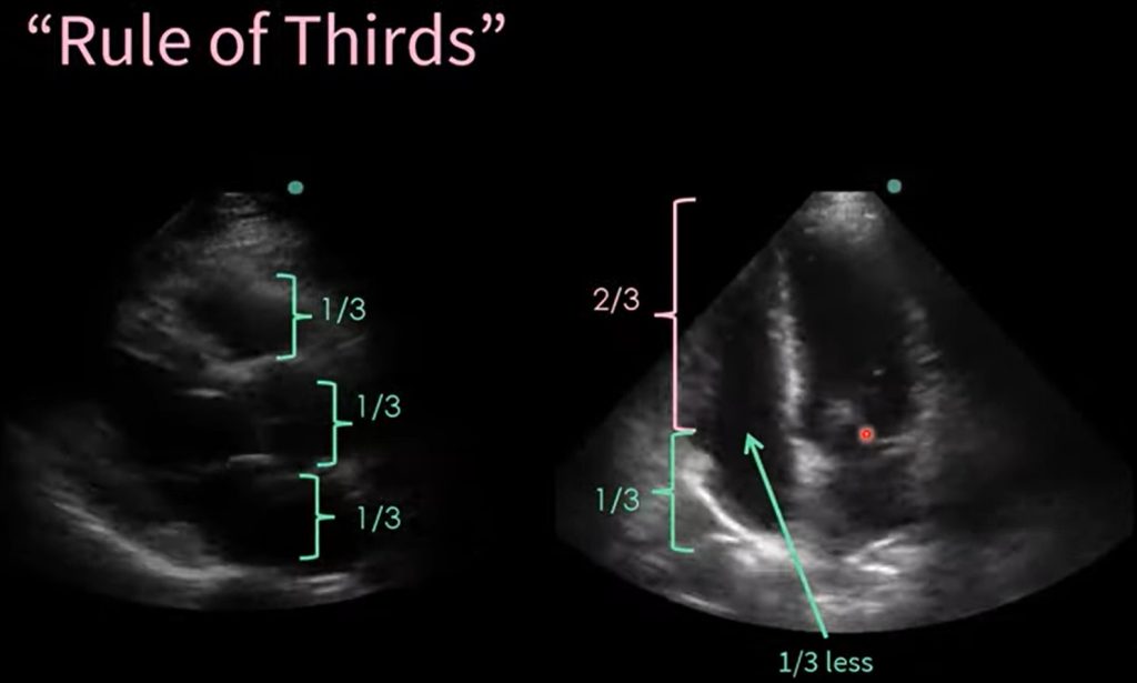
By eyeballing, the sonographer can estimate the normal size by using the 1/3 rule on the Parasternal Long Axis which should be 1/3 left atrium, 1/3 aortic root and 1/3 right ventricle. While using 1/3 rule for size of atrias and 2/3 for ventricle.
Mastering these standard views in echocardiography is essential for performing effective cardiac assessments in clinical and emergency settings. The ability to quickly obtain and interpret these views can significantly impact patient care, allowing for rapid decision-making in acute scenarios.
