6 Basic Principles, Techniques and Protocol of Lung Ultrasound
Fairrul Kadir
Fundamental Principles of Lung Ultrasound
Lung ultrasound (LUS) operates on unique principles due to the distinct nature of lung tissue:
- Ultrasound and Air Interaction:
The primary challenge in lung ultrasound is the high impedance difference between air in the lungs and surrounding tissues. This difference leads to the creation of specific artefacts essential in lung imaging.
- Artefacts as Diagnostic Tools:
Unlike other forms of ultrasound, in LUS, artefacts such as A-lines (horizontal reverberation artefacts) and B-lines (vertical comet-tail artefacts) are key diagnostic elements. These artefacts provide crucial information about the underlying lung pathology.
Lung Ultrasound Techniques
The technique of performing lung ultrasound involves several steps and considerations:
- Patient Positioning:
The patient’s position can vary based on their clinical condition. For critically ill patients, the exam often begins with them in a supine or semi-recumbent position. The probe is moved around to access different lung areas as needed.
- Probe Selection:
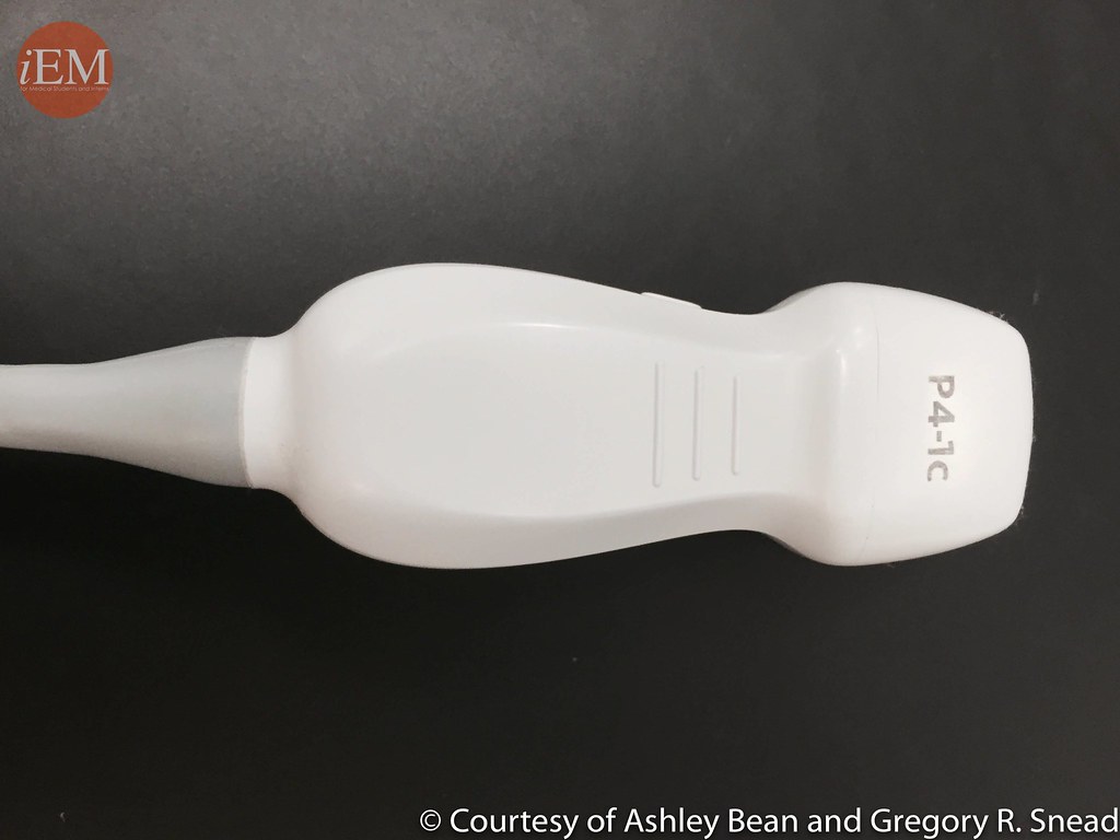
A high-frequency linear probe is typically used for superficial structures and pleural examination. In contrast, a low-frequency curvilinear or phased array probe is preferred for deeper lung tissue examination.
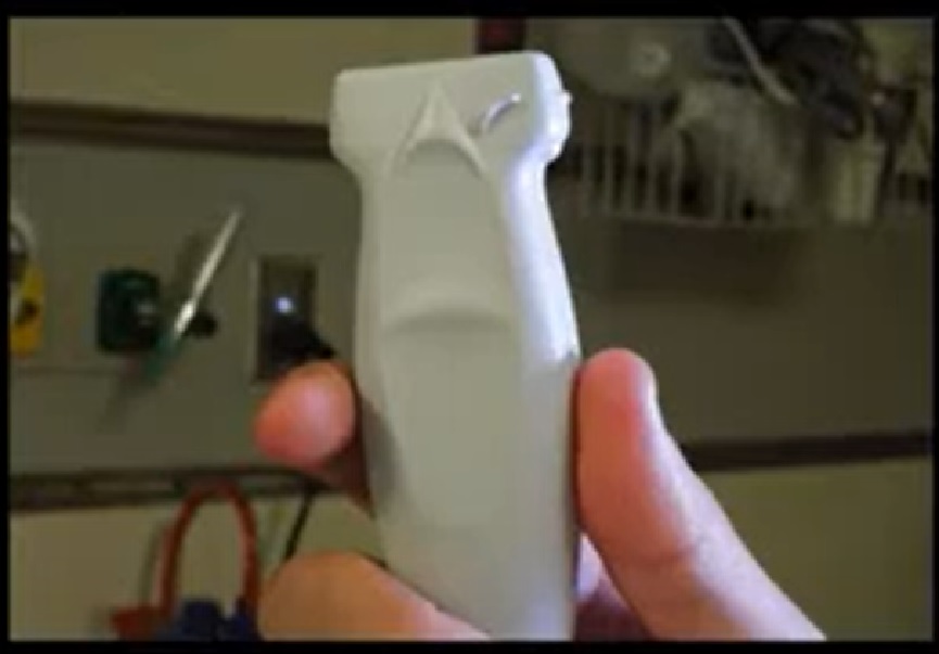
High Frequency Linear Probe. Adapted from “Video 18 Tutorial on ultrasound for DVT” by International Emergency Medicine Education Project is licensed under CC BY 4.0
- Optimal Machine Settings:
Adjusting the ultrasound machine’s settings, such as depth, gain, and focus, is crucial for obtaining clear images. These settings enhance the visibility of lung structures and pertinent artefacts.
- Scanning Methodology:
A systematic approach is vital, ensuring all relevant lung zones are thoroughly examined. The probe is oriented in both longitudinal and transverse planes for comprehensive coverage.
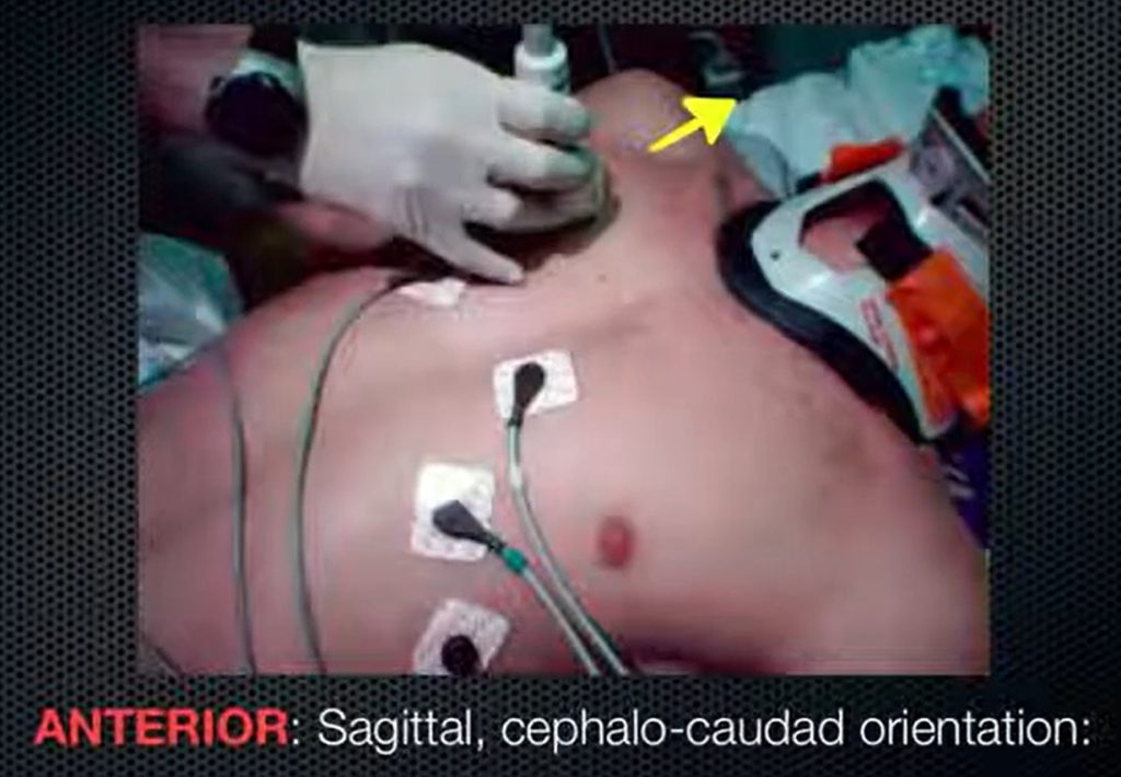
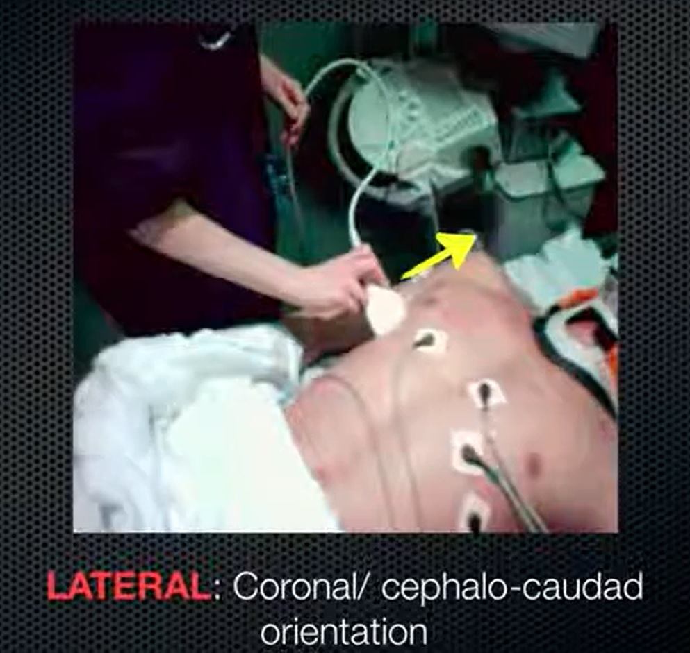
- Standard Lung Views:
The examination generally includes anterior, lateral, and posterior views on both sides of the chest. Each view is assessed systematically for specific artefacts and signs that indicate different lung conditions.
Lung Ultrasound Protocol
- Step-by-Step Examination:The clinician typically starts the examination from the anterior chest and progresses laterally and posteriorly, adjusting the patient’s position as necessary.
- Identification of Landmarks:Recognising anatomical landmarks such as the ribs and pleural lines is essential for orientation and accurate assessment during the scan.
- Interpretation of Findings:Real-time interpretation of the ultrasound findings in the context of the patient’s clinical picture is crucial. Patterns and distribution of artefacts inform diagnoses such as pneumothorax, pleural effusion, or interstitial lung disease.
Probe Positioning in Lung Ultrasound
Getting an adequate and usable view of Lung Ultrasound requires understanding of the lung anatomy. Roughly, a placement of the probes at the anterior chest and lateral chest will give off general view of the lungs. As shown by pictures below.
Systematically, the chest can be divided into 8 zones where probes can be placed, and different zones has its own significance in detecting specific disease findings, as shown in the picture below.
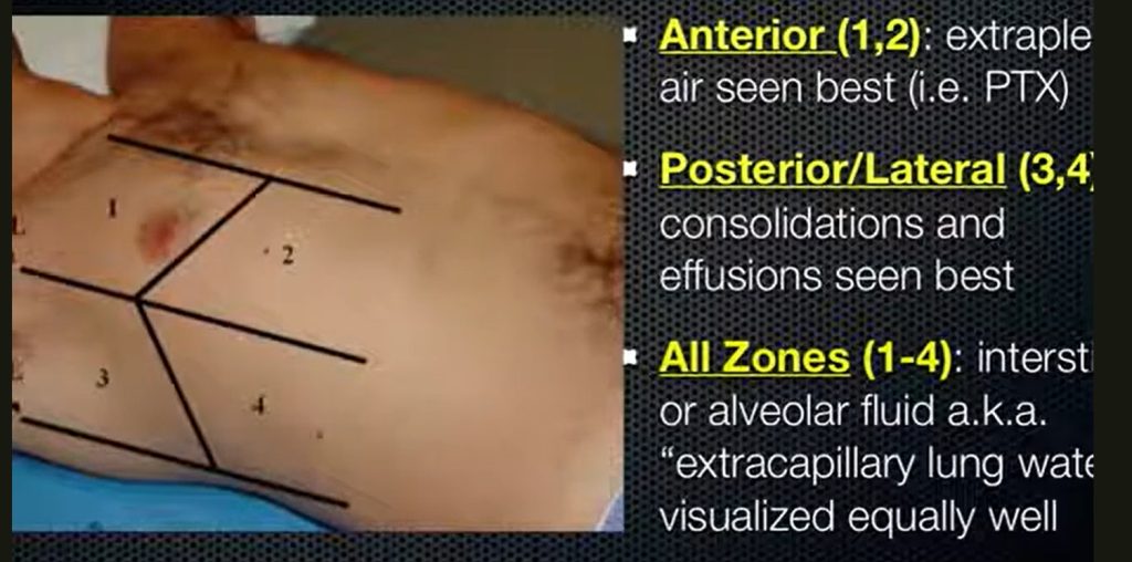
Adapted from “clinical sonography │Lung Ultrasound” by Maladies infectieuses Algerie is licensed under CC BY 4.0
