25 Assessing Basic Cardiac Function and Anatomy
Mexmollen Marcus
Evaluating Left and Right Ventricular Size and Function
- Left Ventricular Size and Function:
“Video 4 Normal Left Ventricular LV contractility” by International Emergency Medicine Education Project is licensed under CC BY 4.0
- Tools: Use the parasternal long axis and short axis views primarily.
- Assessment: Look at the size of the left ventricle at end-diastole and end-systole. Evaluate the thickness of the ventricular walls and the motion of the septum and posterior wall. Measure the ejection fraction (EF), which indicates how well the left ventricle pumps blood with each heartbeat.
- Normal Function: The left ventricle should contract symmetrically and empty effectively, with an EF typically between 55% and 70%.
2. Right Ventricular Size and Function:
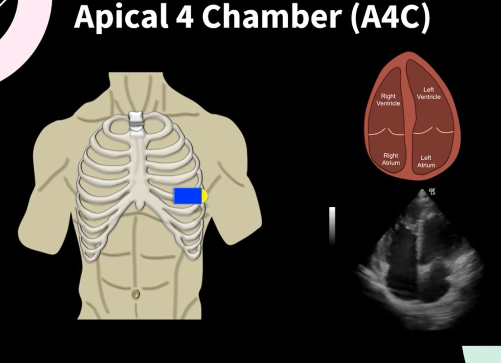
- Tools: The apical four-chamber and subcostal views are best for visualizing the right ventricle.
- Assessment: Observe the size and shape of the right ventricle, which should be smaller than the left and triangular in shape. Check for the movement of the right ventricle wall and its ability to contract.
- Normal Function: Right ventricular function can be more challenging to quantify than left, but general contractility and wall motion are indicative of good function.
Recognizing Signs of Common Pathologies
- Pericardial Effusion:
Adapted from “Pericardial effusion” by International Emergency Medicine Education Project is licensed under CC BY 4.0
- Identification: Look for an echo-free space around the heart, especially noticeable in the parasternal long, apical, and subcostal views.
- Clinical Significance: Significant effusion can lead to cardiac tamponade, a life-threatening condition requiring immediate attention.
2. Cardiomyopathies:
Adapted from “Video 5 Severely decreased LV contractility” by International Emergency Medicine Education Project is licensed under CC BY 4.0
- Types and Signs:
- Dilated Cardiomyopathy: Enlarged ventricular size with decreased systolic function (low EF).
- Hypertrophic Cardiomyopathy: Thickened ventricular walls, especially the septum, with potentially normal EF but impaired filling and outflow.
- Restrictive Cardiomyopathy: Normal ventricle size but stiff walls leading to poor ventricular filling.
- Views for Assessment: All views are useful, but the parasternal and apical views provide the best information on wall thickness and chamber size.
3. Valvular Abnormalities:
Adapted from “Posterior Mitral Valve Prolapse” by Echocardiographer.org is licensed under CC BY 4.0
- Identification: Use primarily the parasternal and apical views to assess the motion and morphology of the valves.
- Common Issues:
- Aortic Stenosis: Thickening of the aortic valve, restricted opening.
- Mitral Regurgitation: Incomplete closure of the mitral valve, seen as backflow during systole in Doppler modes.
- Doppler Ultrasound: Essential for assessing the severity of valvular diseases by measuring the speed of blood flow across the valves.
