1 Ultrasound Basic Physics
Mohammad Firdaus Bolong
Introduction
Ultrasound is a form of mechanical energy that manifests as sound waves with a frequency beyond the range of human hearing (>20,000 Hz). In the medical field, ultrasound technology leverages these sound waves to visualize structures within the body. Understanding the basic physiology of ultrasound is crucial for proper application in clinical settings.
Basic Definitions and Physics
Sound can be heard by human beings in the range of 20 to 20,000 hertz (Hz). For range above that, it will be beyond the human hearing, thus the term “ultrasound”. For diagnostic ultrasound, high frequency is used, ranging from 1 to 15Mega Hz. Different frequencies are used by different transducers in an ultrasound machine for different type of penetration and image acquisition.
Ultrasound wave formed by the transducer uses piezoelectric effect of crystals which are present at the tip of the transducers. When electric current is applied, these crystals are deformed due to its elasticity, which then generates ultrasound waves through the transducer. The reverse effect is achieved when the ultrasound sent from the transducer is reflected back, causing deformation of ghe crystal which will be interpreted as images on the ultrasound scree.
Sound can be described as a mechanical wave resulting from the vibration of particles in a medium (like air, water, or tissue). The basic properties of sound include:
Amplitude: Is peak pressure of wave. Its unit is in decibels (dB) and can describe the loudness of a noise or echo strength in ultrasound. Ultrasound machine probes will be sending sound waves towards a tissue when in use and are able to measure this returned echo once it was reflected back by the tissue, and will appear as images on the screen.
Strong echoes which usually reflected back by dense tissues such as bones will be reflected as bright spots on the screen (hyperechoic) where else weaker echoes returning from less dense tissue such as air or fluids will be reflected as grey areas (hypoechoic) or black (anechoic) on the ultrasound screen.
Frequency (f): Frequency depends on the transducer types. It is important for tissue penetration as well as image resolution. Higher frequency transducer generates better images but have poor penetration, and vice versa. Number of cycles per second, measured in Hertz (Hz).1 cycle per second is 1 Hz.
Speed/Velocity (c): Is the speed of the wave. Velocity is constant in a medium. Distance or depth of a tissue from surface can be calculated using this principle by measuring how long it takes for an ultrasound wave to be reflected back by the scanned medium to the source (ultrasound probe). This principle is also applied by sonars on submarines and also fishing radar.
Wavelength (λ): Distance the waves travels in between peaks in a single cycle. Referring to the formula of (c = f x λ), because velocity is constant, higher frequency will have lower wavelength, which results in decreased penetration.
The relationship between these properties is given by: c=f×λ
This simple equation is best narrated by the video below :
Adopted from “Ultrasound Physics 1 – Sound as Waves” by Marc Kohli is licensed under CC BY 3.0
Interaction of Ultrasound with Tissues
When ultrasound waves encounter tissue, several interactions can occur:
Reflection: Ultrasound waves are reflected to its source, in this case the ultrasound transducer.
Refraction: Redirection of waves crossing a boundary travelling between different mediums. Think of watching a fish in a pond effect, where the fish appears closer to the surface due to refraction of the light waves travelling from air into the water, because propagation speed is different in various mediums.
Attenuation: It is describes as progressive reduction in amplitude as the wave travels through tissue due to absorption, scattering, and reflection, causing the wave to lose energy.
Transmission: Waves pass through the tissue.
The percentage of reflected vs. transmitted energy depends on the acoustic impedance of the tissues. Mismatch in impedance between two media results in more reflection.
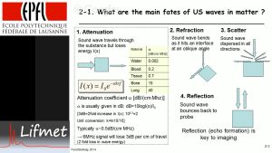
Adopted from “The four Fates of Ultrasound Waves in Tissue” by Fund Biolmag is licensed under CC BY 3.0
1.3 Generation and Detection of Ultrasound
Ultrasound machines generate and detect sound waves using piezoelectric crystals. These crystals:
Convert electrical energy to mechanical vibrations (ultrasound waves) when an electrical signal is applied.
Conversely, generate electrical signals when compressed by mechanical vibrations.
This dual ability allows for both transmission and reception of ultrasound waves.
1.4 Modes of Ultrasound Imaging
A-mode (Amplitude mode): One-dimensional display showing amplitude of the reflected signal vs. depth.
B-mode (Brightness mode): Two-dimensional grayscale image where the brightness represents the amplitude of the reflected signal.
M-mode (Motion mode): Used primarily for cardiac imaging to display the movement of structures over time.
Doppler mode: Assesses the speed and direction of blood flow by measuring the frequency shift of reflected ultrasound waves.
1.5 Safety and Bioeffects
While ultrasound is generally considered safe, it does have bioeffects, mainly due to:
Thermal effects: Absorption of energy can cause localized heating.
Mechanical effects: Include cavitation (formation of gas bubbles) and radiation force.
However, acoustic power, the energy that leaves the transducer is set to default in most ultrasound machines to prevent these effects. This is because of the concept of ALARA (As Low As Reasonably Acceptable), which means to use the lowest energy or radiation possible to obtain the information needed for diagnosis.
Key Takeaways
- It is important to understand the formula of sound wave c=f×λ
- Sound waves react differently when travelling between mediums
- Ultrasound waves are generated through piezoelectric effects
- Different transducers have different frequency which gives varying wave penetrations and image quality
CHAPTER EXERCISE
1. What is the frequency range of sound waves considered as ultrasound?
2. How is an ultrasound image formed?
3. Explain “ALARA” concept
Is a machine that uses ultrasound waves emitted from transducers to view internal organs
Part of an ultrasound machine that generates ultrasound wave that will penetrate tissues and in return will create images on the screen
Crystals embedded in the tip of transducers that deforms on application of electric energy and converts it into ultrasound wave
Sound wave beyond the hearing of humans that is used by ultrasound machine to penetrate tissues and will be reflected back to the tranducers of ultrasound machine
logarithmic unit to measure sound level. Unit (dB)
