7 Key Signs and Interpretations in Lung Ultrasound
Fairrul Kadir
Lung ultrasound (LUS) is characterized by several key signs, each with specific interpretations crucial in diagnosing various pulmonary conditions. Understanding these signs is fundamental to using LUS effectively in clinical practice.
- Bat Sign
- Description: The bat sign is identified as the pleural line with the rib shadows on either side, resembling the wings of a bat. It serves as an important landmark in lung ultrasound, indicating the starting point for scanning.
- Clinical Interpretation: Recognizing the bat sign is essential for proper orientation during lung ultrasound. It helps in differentiating pleural from parenchymal abnormalities and ensures that subsequent findings are accurately located and interpreted.
Bat Sign Appearance in Lung Ultrasound. Adapted from “Video 1 Bat Sign” by International Emergency Medicine Education Project is licensed under CC BY 4.0
- Lung Sliding
- Description:Lung sliding is the to-and-fro movement of the pleural line, visible during respiratory cycles.
- Clinical Interpretation:The presence of lung sliding is a normal finding, indicating the visceral and parietal pleurae are intact and sliding over each other. Its absence can indicate pneumothorax, particularly if a lung point is identified.
Lung Sliding in Lung Ultrasound. Adapted from “Video 2 Lung Sliding” by International Emergency Medicine Education Project is licensed under CC BY 4.0
- A-Lines
- Description:A-lines are horizontal reverberation artefacts that represent normal air-filled lung tissue. They are parallel to the pleura and equidistant from each other.
- Clinical Interpretation:A-lines typically indicate normal aeration of the lungs. However, their presence does not exclude pathology, especially in the context of a symptomatic patient.
A Lines in Lung Ultrasound. Adapted from “Video 5 A Lines” by International Emergency Medicine Education Project is licensed under CC BY 4.0
- B-Lines
- Description:B-lines are vertical, laser-like artefacts that extend from the pleural line to the bottom of the screen without fading. They move synchronously with lung sliding.
- Clinical Interpretation:B-lines are indicative of interstitial syndrome, which can be caused by various conditions such as pulmonary oedema, interstitial pneumonia, or fibrosis. The number and distribution of B-lines help assess the pathology’s severity and extent.
B Lines in Lung Ultrasound. Adapted from “Video 6 B Lines” by International Emergency Medicine Education Project is licensed under CC BY 4.0
- Lung Point
- Description:The lung point is a specific sign where the presence and absence of lung sliding are seen alternatively in the same intercostal space during respiratory cycles.
- Clinical Interpretation:The lung point is pathognomonic for pneumothorax; it marks the boundary between the collapsed and non-collapsed lung.
Lung Point in Lung Ultrasound. Adapted from “Video 4 Lung Point on B Mode” by International Emergency Medicine Education Project is licensed under CC BY 4.0
- Consolidation
- Description:Consolidation appears as a hypoechoic or tissue-like structure within the lung, often with sonographic air bronchograms (dynamic air bronchograms move with respiration).
- Clinical Interpretation:Consolidation typically suggests pneumonia. The size and location of the consolidation can aid in determining the extent of the infection.
Lung Ultrasound in Pneumonia showing consolidation appearance.Adapted from “616 pneumonia” by International Emergency Medicine Education Project is licensed under CC BY 4.0
- Pleural Effusion
- Description:Pleural effusion appears as an anechoic or hypoechoic space between the parietal and visceral pleura.
- Clinical Interpretation:Detecting pleural effusion is essential in diagnosing conditions like heart failure, infections, or malignancies. LUS can also guide thoracentesis.
Pleural effusion detected at right costophrenic angle using Lung Ultrasound. Adapted from “SS Video 22 Small Pleural Effusion” by International Emergency Medicine Education Project is licensed under CC BY 4.0
These key signs form the cornerstone of lung ultrasound diagnostics. Accurate interpretation of these signs requires correlating them with the patient’s clinical presentation and history. For instance, the presence of multiple B-lines in a patient with dyspnea may suggest pulmonary oedema, especially if correlated with clinical signs of heart failure. Similarly, the absence of lung sliding in a trauma patient may prompt an urgent evaluation for pneumothorax.
In clinical practice, lung ultrasound is about recognizing these signs and their implications in the broader context of patient care. It enables clinicians to make rapid bedside assessments that significantly impact patient management and outcomes, especially in emergency and critical care settings.
Clinical Applications of Lung Ultrasound
Lung ultrasound (LUS) has become invaluable in various clinical settings due to its versatility, non-invasiveness, and immediate diagnostic capabilities. Its applications span different medical disciplines, significantly impacting managing patients with various pulmonary conditions.
- Diagnosis of Pneumothorax
- Application:LUS is highly effective in detecting pneumothorax, especially in emergency and critical care settings. The absence of lung sliding, B-lines, and the presence of a lung point are key indicators.
- Diagnosis of Pneumothorax with M-mode Ultrasound:
- Sea-Shore Sign:
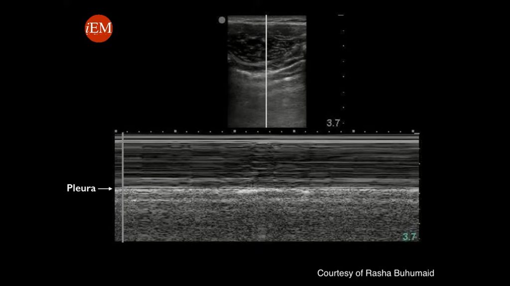
Seashore sign which signifies normal lung finding in M Mode in Lung Ultrasound. Adapted from “Video 14 Tutorial Lung ultrasound for pneumothorax” by International Emergency Medicine Education Project is licensed under CC BY 4.0
In M-mode, a normal lung exhibits the ‘sea-shore sign.’ The static chest wall appears as a granular pattern (representing the ‘sand’) above the pleural line, while the sliding lung beneath the pleural line creates a horizontal, linear pattern (representing the ‘sea’). This sign indicates normal lung sliding and the absence of pneumothorax.
- Barcode Sign:
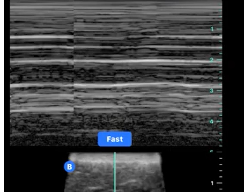
Barcode sign in M Mode during Lung Ultrasound which can signifies pneumothorax. Adapted from “Lung US – absence of lung sliding and comet tail artefacts – pneumothorax and barcode sign” by International Emergency Medicine Education Project is licensed under CC BY 4.0
In the presence of pneumothorax, the ‘barcode sign’ (also known as the ‘stratosphere sign’) is observed. This pattern shows a continuous granular or linear pattern throughout, without the distinct separation seen in the sea-shore sign. It indicates the absence of lung sliding, which is highly suggestive of pneumothorax.
- Advantages:It provides a rapid and reliable diagnosis, which is crucial for timely intervention in life-threatening situations like trauma or post-procedural complications.
- Assessment of Pulmonary Edema
- Application:LUS is used to identify and quantify pulmonary oedema. The presence and number of B-lines can indicate the severity of interstitial fluid.
- Advantages:This is particularly beneficial in differentiating cardiac from non-cardiac causes of dyspnea, guiding fluid management in heart failure patients, and monitoring response to treatment.
- Evaluation of Pleural Effusion
- Application: LUS effectively detects pleural effusions, characterises them as transudative or exudative, and guides pleural fluid aspiration.
- Advantages: It offers a safer and more accurate approach than traditional methods, reducing the risk of complications associated with blind thoracentesis.
- Diagnosis of Pneumonia
- Application: LUS can identify lung consolidation characteristics of pneumonia and help differentiate it from other causes of acute respiratory distress.
- Advantages: It is particularly useful in settings where traditional imaging modalities are not feasible, like in pediatric populations or resource-limited environments.
- Guiding Critical Care Management
- Application: In critically ill patients, LUS aids in rapidly assessing acute respiratory failure, guiding ventilator management, and monitoring lung recruitment manoeuvres.
- Protocols: The BLUE (Bedside Lung Ultrasound in Emergency) protocol is utilized for rapid assessment of respiratory failure, and the FALLS (Fluid Administration Limited by Lung Sonography) protocol helps manage circulatory failure.
- Applications in Cardiology
- Application: Cardiologists use LUS to evaluate pulmonary congestion in heart failure patients, aiding in diagnosing and managing acute decompensated heart failure.
- Advantages: It complements echocardiographic findings, providing a more comprehensive cardiac-pulmonary assessment.
- Applications in Pediatrics
- Application: In pediatric care, LUS is used for diagnosing pneumonia, monitoring chronic lung diseases, and evaluating neonatal respiratory conditions.
- Advantages: Its radiation-free nature makes it particularly suited for pediatric patients, minimizing the risks associated with repeated imaging.
