20 Anatomy and Physiology of the Aorta
Lo Zhen Zhen
Overview of Aortic Structure
The aorta, which originates from the heart’s left ventricle, is the largest artery in the body. It is divided into ascending aorta, aortic arch, descending thoracic aorta, and abdominal aorta. The aorta wall is made up of three layers:
- Intima: The innermost layer in direct contact with the blood flow.
- Media: The middle layer, composed of smooth muscle and elastic tissue, provides strength and flexibility.
- Adventitia: The outermost layer, which provides extra support and structure.

Layers of Normal and Dissected Artery. Adapted from “Arterial dissection.webp” by Nadezdha D. Kiriyak is licensed under CC BY 4.0
Understanding these layers is crucial in ultrasound as variations and pathologies often involve specific layers, such as dissections occurring within the media.
Branches of the Aorta
The aorta gives off several major branches, which supply blood to the head, arms, and major organs. Key branches include:
- Branches of the Ascending Aorta: The coronary arteries.
- Branches of the Aortic Arch: The brachiocephalic trunk, left common carotid, and left subclavian arteries.
- Branches of the Descending Thoracic Aorta: Intercostal arteries.
- Branches of the Abdominal Aorta: Including the celiac trunk, renal arteries, and iliac arteries.
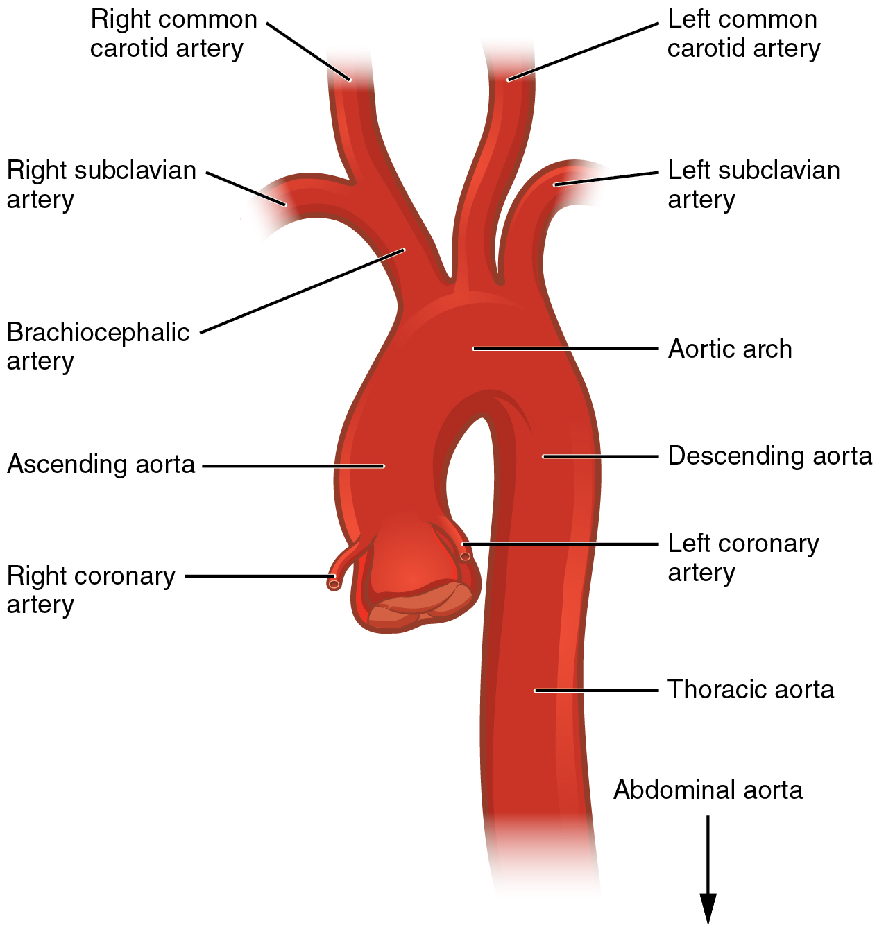
Branches of the aorta. Adapted from “2121 Aorta.jpg” by OpenStax College is licensed under CC BY 3.0
These branches serve as landmarks for orientation during ultrasound scans and can also be disease locations, such as aneurysms or stenosis.
